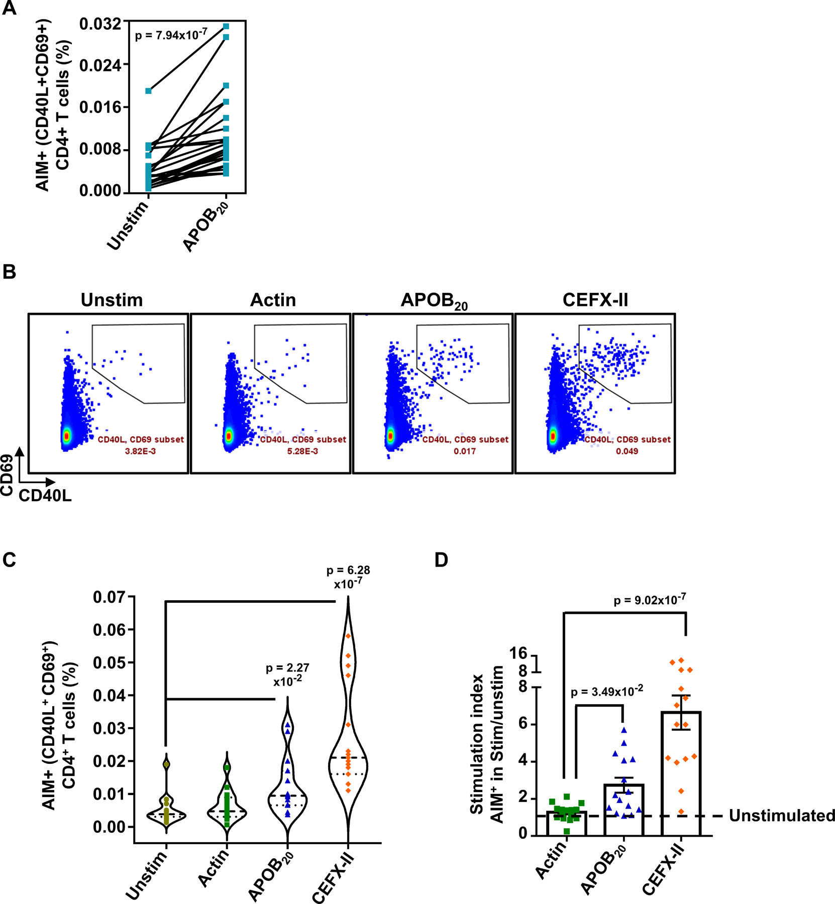
Figure 2: Activation-induced surface expression of CD40L and CD69 (AIM assay) allows sensitive detection of APOB-specific CD4+T cells in ex vivo stimulated PBMCs.

A) %CD40L+CD69+ (AIM+) CD4+T cells in paired sets of APOB20 stimulated and unstimulated PBMCs. 21 independent donors. B) Representative FACS plots and C) Violin plots showing median frequencies of %CD40L+CD69+ (AIM+) CD4+ T cells in unstimulated and 24h Actin pool or APOB20 pool or CEFX-II pool-stimulated PBMCs. D) Average stimulation indices (%CD40L+CD69+ CD4 T cells in stimulated sets over those in unstimulated control) for Actin, APOB20 and CEFX-II peptide pools. 15 independent donors. Colored symbols represent data from individual donors. Pairwise statistical comparisons (A) were performed with the Wilcoxon test. Statistical tests for mean response across different stimulation conditions and unstimulated (C) or actin (D) sets were performed using Kruskal-Wallis test with Dunn’s multiple comparison testing..
