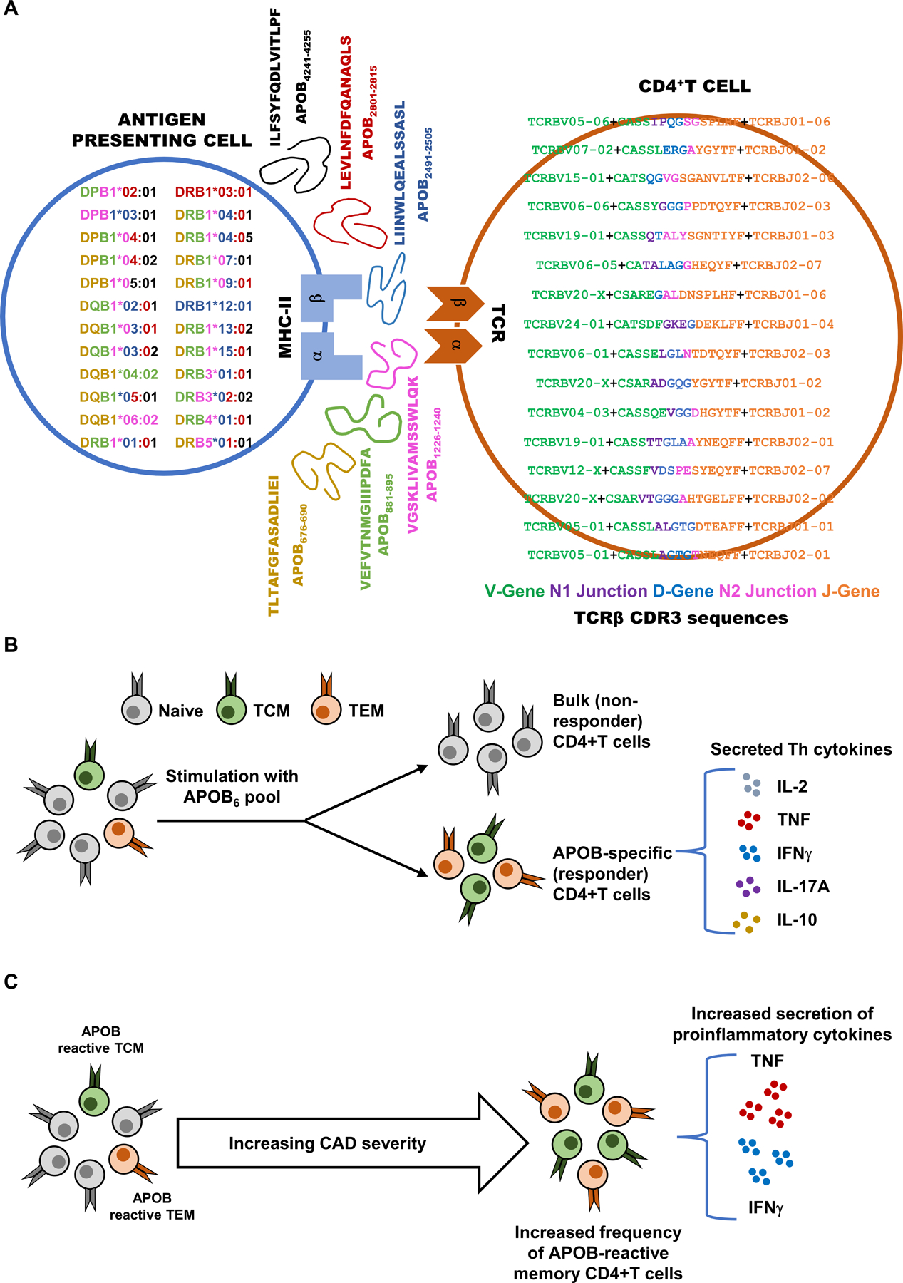
Figure 8: Broadly binding MHC Class-II-restricted immunodominant epitopes in human APOB allow phenotypic evaluation of atherosclerosis-related CD4+T responses and enable identification of APOB-specific autoreactive TCR clones.

A) Human HLA-II binder alleles expressed in donor samples are shown in the antigen presenting cell (left). Each allele is color coded by the corresponding color of the APOB epitopes (middle, with sequence and position in APOB) that it binds. Bio-identities of clones whose AIM+ vs AIM− log10 odds ratios are > 1 and FDR corrected Fisher’s exact test p values < 10−200 are shown in the CD4+ T cell. Sequences are color coded by component. Resolved V gene green, translated CDR3 region (which includes the start of CDR3 encoded by the V gene green, N1 junction purple, D-gene blue, N2 junction pink, end of CDR3 in J gene orange) and resolved J-gene orange are shown. B) Stimulation of human PBMCs with a pool of dominant epitopes (APOB6) elicits memory CD4+T responses and triggers secretion of multiple T helper cytokines. C) Higher frequencies of antigen-experienced APOB6-reactive CD4+T cells and increased proinflammatory cytokine responses are observed in patients with more severe CAD.
