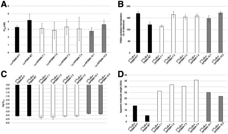
FIGURE 3.
(A) Binding affinities (half-maximal inhibitory concentration [IC50; nM], 1 h, 4°C) of [177Lu]Lu-rhPSMA-7.1 to -7.4 (white; n = 3), [177Lu]Lu-rhPSMA-10.1 and -10.2 (black/white stripes; n = 3), and references [177Lu]Lu-PSMA-617 and [177Lu]Lu-PSMA-I&T (black; n = 3). (B) PSMA-mediated internalization of [177Lu]Lu-rhPSMA-7.1 to -7.4 (white; n = 3), [177Lu]Lu-rhPSMA-10.1 and -10.2 (black/white stripes; n = 3), and references [177Lu]Lu-PSMA-617 and [177Lu]Lu-PSMA I&T (black; n = 3) by LNCaP cells (1 h, 37°C) as percentage of reference ligand ([125I]IBA)KuE). (C) Lipophilicity of [177Lu]Lu-rhPSMA-7.1 to -7.4 (white; n = 6), [177Lu]Lu-rhPSMA-10.1 and -10.2 (black/white stripes; n = 6), and references [177Lu]Lu-PSMA-617 and [177Lu]Lu-PSMA I&T (black; n = 6), expressed as partition coefficient (log D7.4 in n-octanol/phosphate-buffered saline, pH 7.4). (D) AMW of [177Lu]Lu-rhPSMA-7.1 to -7.4 (white), [177Lu]Lu-rhPSMA-10.1 and -10.2 (black/white stripes), and references [177Lu]Lu-PSMA-617 and [177Lu]Lu-PSMA I&T (black), as determined by HSA-mediated size-exclusion chromatography.

