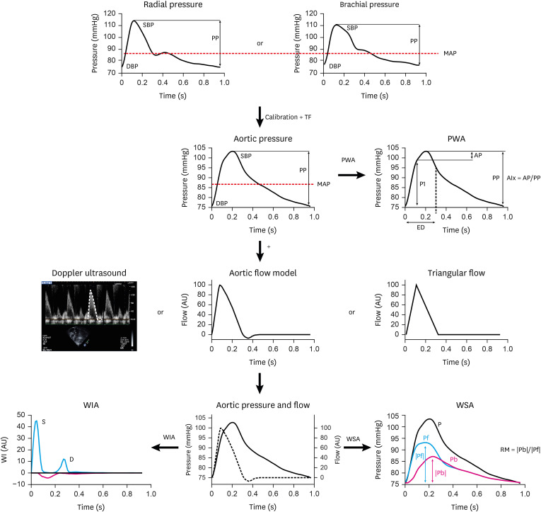
Figure 1. Assessment of pulsatile hemodynamics - overview. Top line: Recording of signal-averaged radial or brachial pressure waveforms with tonometry or brachial cuff. Second line: Following calibration with brachial pressures, aortic waveforms are calculated with a TF. Pulse waveform analysis, based on pressure signals alone, yields measures of the first (P1) and second (P2) systolic peaks for computation of augmented pressure and augmentation index (AP, AIx). Third line: Flow waveforms are obtained, either with Doppler recording of LV outflow (which equals aortic inflow), or as model-deriven flow or triangular flow as a proxy. Bottom line: Combined and time-aligned analysis of a pressure-flow pair is used for wave separation analysis, wave intensity analysis, and other analytical approaches.
Modified from Parragh et al.26) and Hametner et al.,24) and reprinted with permission from Weber and Chirinos.25)
AIx = augmentation index; AP = augmented pressure; DBP = diastolic blood pressure; MAP = mean arterial pressure; Pb = amplitude backward wave; Pf = amplitude forward wave; PP = pulse pressure; PWA = pulse waveform analysis; RM = reflection magnitude; SBP = systolic blood pressure; TF = transfer function; WIA = wave intensity analysis; WSA = wave separation analysis.

