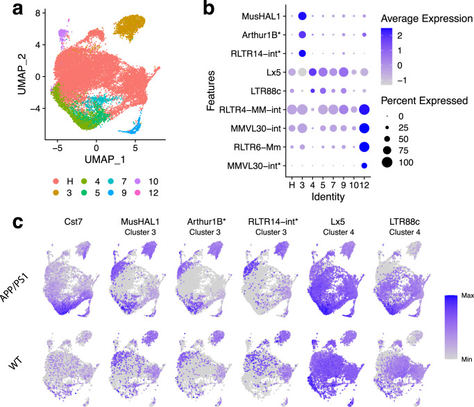
Fig. 6. TE expression detected with SoloTE in the APP/PS1 Alzheimer’s disease mouse model.
a UMAP plot indicating the different cell types (“H”: homeostatic cells). b Dot plot depicting marker TEs per cell types (indicated on the x axis), having a disease-specific effect. c UMAP plots of Cst7 (known gene with increased expression in the disease) and selected TEs. Upper row corresponds to the expression in the AD APP/PS1 samples, whereas the lower row corresponds to the expression in the wild-type (WT) samples. Label below the TE identifier indicates the cluster at which the TE has increased expression. “*” denotes locus-specific TEs.

