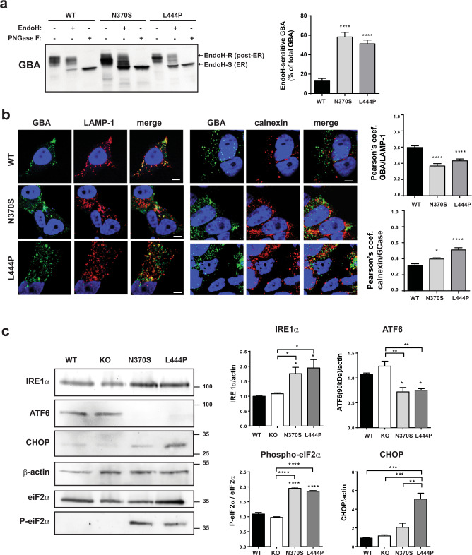
Fig. 2. Mutant GBA proteins are retained in the ER.
a Left: representative immunoblots of GBA before and after treatment with EndoH and PNGase F (as a positive control). b Right: quantification of GBA EndoH-sensitive (EndoH-S) percentage of total GBA protein, n = 8 independent experiments. Representative images and quantification of the colocalization of GBA with LAMP-1 (lysosomes) and calnexin (ER) determined by immunofluorescence. Colocalization was represented as Pearson’s coefficient (fraction of LAMP-1 or calnexin overlapping GBA), scale bar = 5 µM, n > 20 cells/condition. c Representative immunoblots and quantification of ER stress markers (IRE1α, ATF6, phosphorylated eIF2a, and CHOP), n = 3 independent experiments. In all panels data are presented as mean ± s.e.m, statistical significance was established at *p < 0.05, **p < 0.01, ***p < 0.001, ****p < 0.0001 compared to WT samples or between mutant lines when indicated after one-way ANOVA followed by Tukey’s multiple comparisons test.

