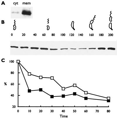
FIG. 1.
The activity of McpA proteolysis is modulated during the cell cycle. (A) Membrane localization of overexpressed McpA. cyt, cytoplasmic fraction; mem, membrane fraction. C. crescentus extracts were separated into cytoplasmic and membrane fractions as described by Shaw et al. (29). (B) Cell cycle immunoblots of the C. crescentus strain MRKA828, which overexpresses McpA constitutively throughout the cell cycle in M2X medium. The numbers above the cell cycle immunoblot represent the time in minutes at which samples were taken, and the C. crescentus drawings denote the progression through the cell cycle. Cell cycle progression was monitored by microscopic analysis. Equal amounts of protein were loaded in each lane. (C) A synchronized culture was labeled with 20 μCi of Tran35S-label (ICN)/ml for 4 min at 0 min (open boxes) or at 40 min (filled boxes) and then chased with 0.2% (wt/vol) Bacto Peptone, 0.1% (wt/vol) Bacto yeast extract, 0.5 mM methionine, and 0.05 mM cysteine. Samples were taken every 10 min except for the last time point. Equal counts were immunoprecipitated with McpA1 antisera. After sodium dodecyl sulfate-polyacrylamide gel electrophoresis, proteins were visualized by a Molecular Dynamics phosphorimager and quantified with IPLabScan software.

