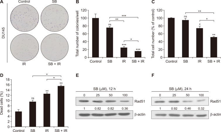
Figure 1. Combinatorial effects of a low dose of SB and IR on clonogenic potential and proliferation of DU145 cells.
(A) Human PCa DU145 cells were seeded in a 6-well culture plate at a density of 600 cells/well and treated with either SB (25 µM) or IR (5 Gy) or in combination and were maintained in a humidified CO2 incubator. After 10 days, plates were processed for the clonogenic assay as described in MATERIALS AND METHODS. Representative images for each treatment group and (B) quantitative data are represented as the total number of colonies/well. (C, D) Fourty thousand cells/well seeded in a 12-well plate were treated with SB (25 µM) and IR (5 Gy). After the 48-hour treatments, cells were trypsinized, harvested and processed for trypan blue staining and live and dead cells were counted using haemocytometer. (E, F) At ~70% confluency, DU145 cells were treated with SB (25, 50, and 100 µM) and harvested after 12 and 24 hours. Whole cell lysates were prepared as described in MATERIALS AND METHODS, and immunoblotting was done for Rad51 protein expression and β-actin was used as loading control. Data are presented as mean ± SE of triplicate samples for each treatment. Results are representative of three sets of independent experiments. Gy, gray; SB, silibinin; IR, ionizing radiation; PCa, prostate cancer; SE, standard error; ns, not significant. *P < 0.05, **P < 0.01, ***P < 0.001.

