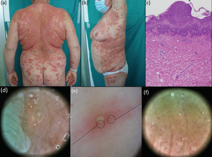
FIGURE 1.

Clinical appearance at presentation time, 26 days after starting hydroxychloroquine therapy, exhibiting lesions in different stages: numerous tiny pustules pustular surrounded by an erythematous halo (e, polarized dermoscopy 17×) evolved into targetoid lesions with raised outer ring that coalesced into wide plaques on the whole trunk (a,b) and remained sparse on the extremities (b). Polarized dermoscopy 30× highlights the presence of clods (asterisks) and curved vessels (arrowheads) along the pustules margin (d) and all over the erythematous halo around the pustule (f). Biopsy specimen from the right side revealed an intraepidermal pustule, lympho‐histiocytic infiltrates in the upper dermis, with dilated papillary capillaries and focal oedema, in absence of sign of vasculitis; focal exocytosis of neutrophils were also present near to the pustule [c, Haematoxylin–eosin, 50×].
