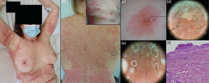
FIGURE 2.

Clinical appearance of GPFE rash occurred 22 days after starting hydroxychloroquine therapy in a 50‐year‐old woman, involving the trunk and proximal extremities with the skin fold sparing. Numerous primary lesions (b, square) consisting of a millimetric pustule surrounded by an erythematous halo (c, polarized dermoscopy 17×) evolved into targetoid lesions with raised pustular borders and merged to form wide plaques (a,b). Polarized dermoscopy 30× reveals clods (asterisks) and curved vessels (arrowheads) along the pustules margin (d) and between pustules (e). Histopathological examination revealing subcorneal pustule with focal spongiosis and acanthosis with focal exocytosis of neutrophils; the upper dermis shows dilated capillaries, oedema, lympho‐histiocytic infiltrates with numerous neutrophilic granulocyte and occasional eosinophilic granulocytes [f, Haematoxylin–eosin, 50×].
