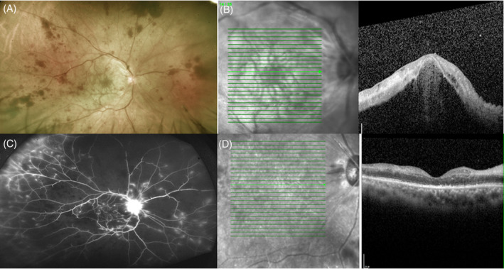
FIGURE 1.

Combined central retinal artery/vein occlusion and exudative retinal detachment of right eye after COVID‐19 vaccination. (A) Fundoscopic examination showed extensive retina hemorrhages, attenuated artery, and whitening of macular area. (B) Optical coherence tomography (OCT) demonstrated retinal detachment and hyper‐reflective change of inner retina. (C) Late‐phase fluorescein angiography showed wide non‐perfused area and peripheral vessel as well as disc fluorescein leakage. (D) 1 month after the treatment, OCT revealed subsided subretinal fluid, but a disorganized and atrophic retina structure
