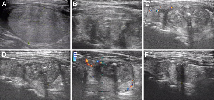
Figure 3.
Imaging of a 63-year-old woman with Bethesda IV nodules who underwent microwave ablation (MWA). (A) An isoechoic solid thyroid nodule was detected in the left lobe with an initial volume of 14.29 ml. (B) Ultrasound examination showed enlarged ablation area (20.16 ml) immediately after MWA. (C–F) The volume of the ablation area was 12.39, 6.33, 3.13, and 2.04 ml at 1, 3, 6, and 12 months after MWA, respectively.

