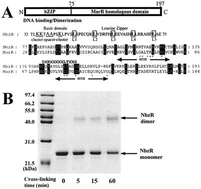
FIG. 4.
(A) Schematic domain structure of NbzR. The typical characteristics of bZIP, repeating leucine residues and a preceding basic region, and an alignment of NbzR (residues 77 to 193) with MarR from E. coli K-12 (GenBank accession no. AE000250) are shown below. A preliminary consensus sequence (DXRXXXXXLTXXG) of the MarR family (47) is shown above the alignment. The putative HTH regions in MarR are indicated below the alignment. Amino acids highlighted in black boxes are identical residues. #, Residues of MarR causing negative complementary trans-dominant mutations; ∗, residue of MarR making a specific operator contact. (B) Chemical cross-linking of NbzR by EDC. NbzR (1 mg/ml) was reacted with 2 mM EDC and 5 mM NHS at 4°C for different times as described in Materials and Methods. Arrows indicate monomeric and dimeric forms of NbzR.

