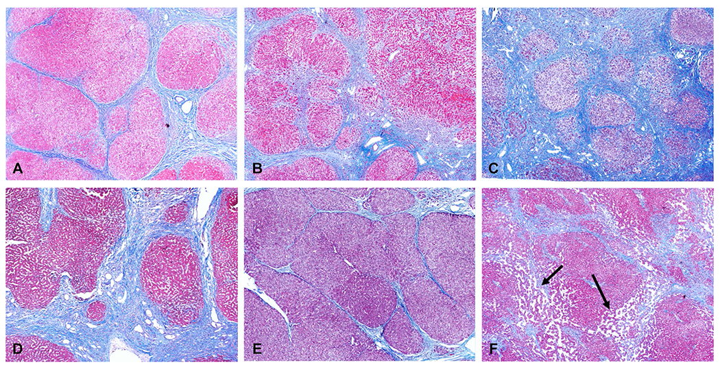
Fig. 3.

Examples of cirrhosis from chronic hepatitis C (A), NASH-non-alcoholic steatohepatitis (B), ASH-alcoholic steatohepatitis (C), primary sclerosing cholangitis (D), auto immune hepatitis (E) and cardiac cirrhosis (F). Note the different degrees of fibrosis deposition. No hepatic steatosis is seen in both NASH and ASH examples which is common at this stage of chronic liver disease. Dilated sinusoidal spaces (arrows, F) are seen secondary to chronic venous outflow obstruction in this case of cardiac cirrhosis
