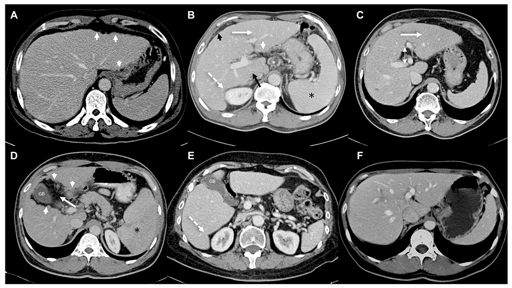
Fig. 5.

Contrast enhanced CT images from different patients illustrating the morphological changes in liver in chronic liver disease. Nodular outline of the liver (short arrows in A, B, and D), atrophic right lobe with enlarged caudate lobe (black arrow in B), enlarged left lobe (white arrow in B and C), increased abdominal fat or creeping fat sign (short black arrow, B), increased periportal space (double headed arrow, C), enlarged gall bladder fossa sign (white arrow, D), posterior hepatic notch sign (broken white arrow, B and E). Note splenomegaly (asterisk, B and D) consistent with portal hypertension. Compare with normal appearance of liver in a chronic hepatitis B patient (F) with biopsy proven stage 2 fibrosis. Morphological changes are usually seen in advanced fibrosis or cirrhosis and mostly absent in early fibrosis stages. GB = gall bladder
