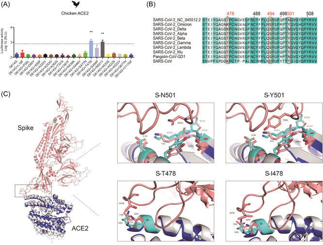
Figure 2.

The characteristic of SARS‐CoV‐2, T478I, and N501Y mutations. (A) Hela cells expressing chicken ACE2 were infected by 20 SARS‐CoV‐2 spike mutations pseudoviruses. At 48 h postinfection, pseudovirus entry efficiency was determined by measuring luciferase activity in cell lysates. The results were presented as the mean relative luminescence units (RLUs) and the error bars indicated the logarithm (base 10) of the standard deviations of the RLUs (n = 9). (B) Sequence alignment of the RBD sequences from the SARS‐CoV‐2, SARS‐CoV‐2 variants, and SARS‐CoV. (C) Structural models of N501Y and T478I were generated based on the crystal structure of the SARS‐CoV‐2 S/ACE2 complex. SARS‐CoV‐2 S, chicken ACE2, and human ACE2 were colored in pink, gray, and blue, respectively. Residues involved in the interaction are labeled. ACE2, angiotensin‐converting enzyme 2; RBD, receptor‐binding domain.
