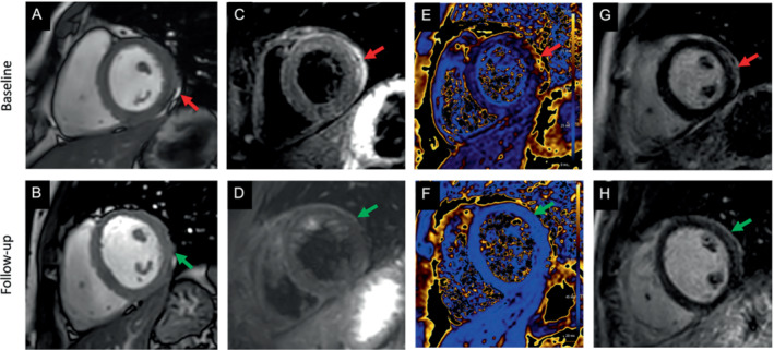
Figure 3.

Cardiac magnetic resonance (CMR) images of a patient with signs of a myopericarditis after mRNA SARS‐CoV‐2 vaccination with Spikevax (Moderna). Full description of this case can be found in Jahnke et al. 65 One day after vaccination the patient complained about chest pain and discomfort, shortness of breath, limited physical capacity and malaise. High‐sensitivity troponin T was elevated up to 526 ng/L (normal <14 ng/L). C‐reactive protein, N‐terminal pro‐B‐type natriuretic peptide, electrocardiogram, echocardiography, coronary angiogram and computed tomography pulmonary angiography were normal. CMR was normal for function (including strain) (A), but demonstrated slight pericardial effusion (red arrow in A). T2 weighted images indicated a regional oedema anterolateral/inferolateral (basal) with corresponding elevated quantitative myocardial T2‐mapping parameters up to 70 ms (normal up to 51 ms at 3 Tesla) (C, E – red arrows). Patchy subepicardial late gadolinium enhancement (LGE) indicating inflammatory myocardial necrosis (G). Pericardial enhancement in the T2‐weighted and LGE images in corresponding locations indicated a pericardial involvement as well (C, E). The findings resolved at 4‐month CMR follow‐up (green arrow in B, D, F, H).
