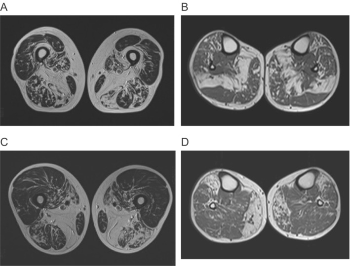
Figure 2.

Magnetic resonance imaging (MRI) of F1.III:12 (A‐B) and F1.IV:4 (C‐D). F1.III:2, at age 48 years, showed severe fatty involvement of vastus intermedius and adductor magnus on the thigh with milder fatty involvement in all hamstrings, adductor longus, and vastus medialis (A). The soleus is severely involved with milder changes in tibialis anterior and medial gastrocnemius (B). In the thigh of F1.IV:4 adductor magnus is replaced with milder fatty involvement of hamstrings, vastus medialis, and sartorius (C). On the lower legs, the medial gastrocnemius is asymmetrically, while the tibialis anterior is more symmetrically involved (D).
