Figure 3.

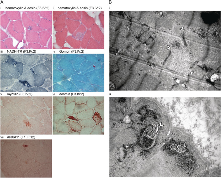
(A) Histochemical and immunohistochemical stainings of left deltoid muscle biopsy sections from F3.IV:2. Hematoxylin & eosin (HE) shows rimmed vacuolated fiber (i) and a larger area of myofibrillar disarray (ii). NADH‐TR staining shows irregular internal architecture with focal areas lacking oxidative activity (iii). Gomori's trichome staining shows red‐purple cytoplasmic inclusions (iv). Abnormal protein accumulations are stained with myotilin (v), larger myofibrillar disarrays with desmin (vi). Staining with anti‐annexin A11 shows positive intravacuolar accumulations (vii). (B) Ultrastructural findings in the muscle biopsy consist of (i) myofibrillar abnormalities with disorganization of the sarcomeric structure and Z‐disc dissolution (shown with white arrowhead), and (ii) subsarcolemmal autophagic material with myeloid formations (shown with white arrowhead).
