Figure 2.
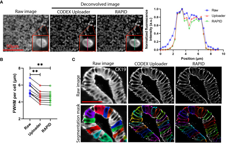
3D image deconvolution improves image contrast in CODEX data. (A) Representative raw and deconvolved images of a cervical lymph node, and plot of normalized fluorescence intensity versus lateral position of a selected cell corresponding to the raw and deconvolved images. (B) Quantification of the FWHM for signals cross ten randomly selected cells. (C) Representative raw and deconvolved images of cytokeratin 19-stained pancreatic ducts and corresponding predicted whole-cell segmentation mask. Wilcoxon matched-pairs signed rank test was used in (B). **P < 0.01.

