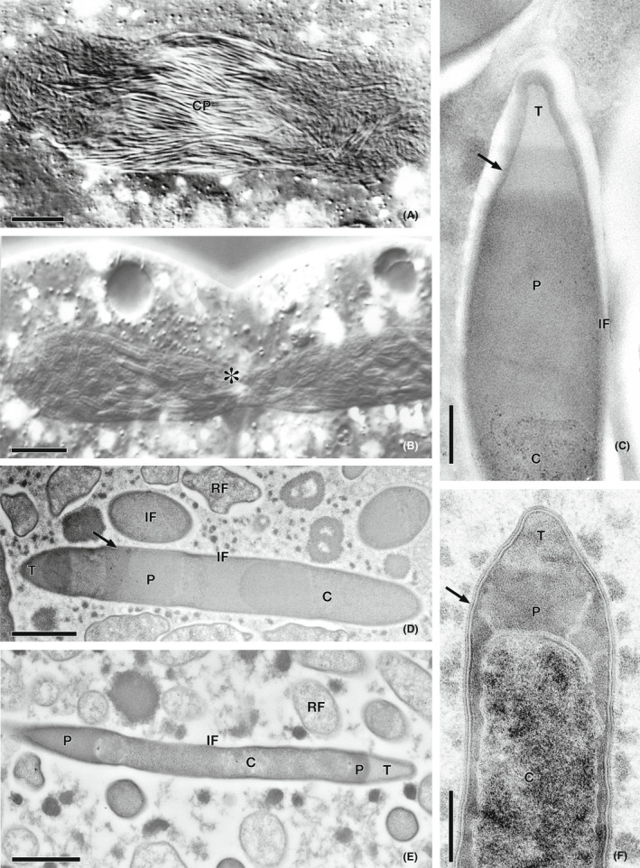
FIGURE 1.

Morphology of Holospora and Holospora‐like bacteria. (A) Equatorial connecting piece (CP) of a dividing Paramecium caudatum macronucleus infected with H. obtusa. (B) Macronucleus of P. putrinum infected with “Ca. Gortzia yakutica” undergoing division shows the absence of the CP (asterisk). (C) Ultrastructure of the infectious form (IF) of “Ca. Holospora parva”; recognition tip (T); the arrowhead indicates the subdivision of periplasmic space (P); bacterial cytoplasm (C). (D) “Ca. Gortzia infectiva” reproductive form (RF), other abbreviations are the same. (E) IF and RF of Preeria caryophila. IF with two recognition tips, located at the opposite ends of the bacterial cell. (F) IF of “Ca. Gortzia shahrazadis”. All abbreviations are the same as in (C–E). (A, B) differential interference contrast microscopy; (C–F) transmission electron microscopy. Scale bars represent 20 μm (A), 10 μm (B), 0.3 μm (C), 1.0 μm (D), 0.6 μm (E), 0.5 μm (F)
