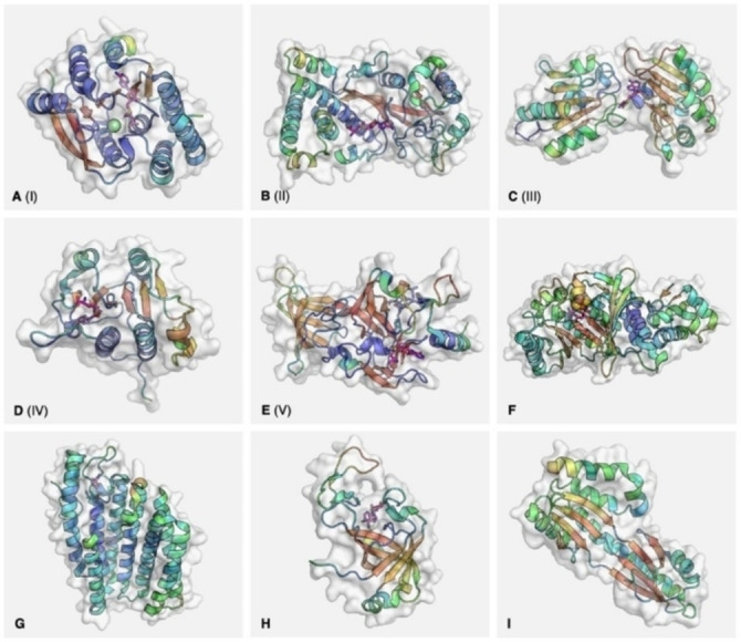
Figure 1.

Representative structures for the known classes of methyltransferase. PDB codes: A – 6LFE (catechol‐O‐methyltransferase); B – 1MSK (methionine synthase, reactivation domain); C – 1CBF (cobalt precorrin‐4‐methyltransferase); D – 1MXI (tRNA (cytidine(34)‐2’‐O)‐methyltransferase); E – 1O9S (SET7/9); F – 3RFA (RlmN); G – 5VG9 (isoprenylcysteine carboxyl methyltransferase); H – 2NV4 (AF0241); I – 1TLJ (Taw3). Secondary structures represented within the molecular surface as cartoons, coloured by B factor: blue (high) to red (low) for α‐helices, the reverse for β‐sheets. Cofactor represented as magenta sticks. For E (1O9S), histone peptide represented as blue sticks. For F (3RFA), iron‐sulfur cluster represented as spheres.
