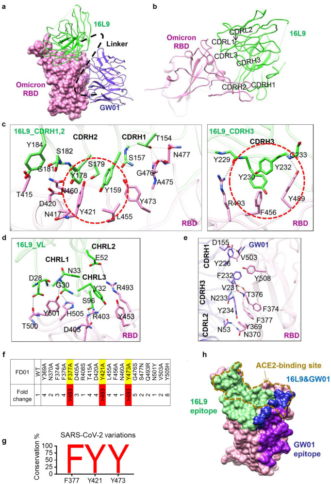
Fig. 5. Two conserved epitopes recognized by FD01.
a Close-up view of the interaction between FD01 and Omicron. The Omicron S RBD is displayed in pink surface. 16L9 and GW01 are shown as cartoons colored green and medium-blue, respectively. b The interactions between S RBD and 16L9. c The detailed interaction between RBD and the heavy chain of 16L9. The residues involved in interactions are represented as sticks. The polar interactions are indicated as dotted lines. The hydrophobic interactions are highlighted by red circles. d The detailed interaction between RBD and the light chain of 16L9. e The detailed interaction between RBD and GW01. f Epitope mapping of FD01. g Conservancy analysis of FD01 epitope. h Surface representation of RBD showing the buried binding site, including 16L9 (green), GW01 (purple), and 16L9 epitope overlapping with GW01 region (blue); goldenrod dotted line indicates receptor-binding site.

