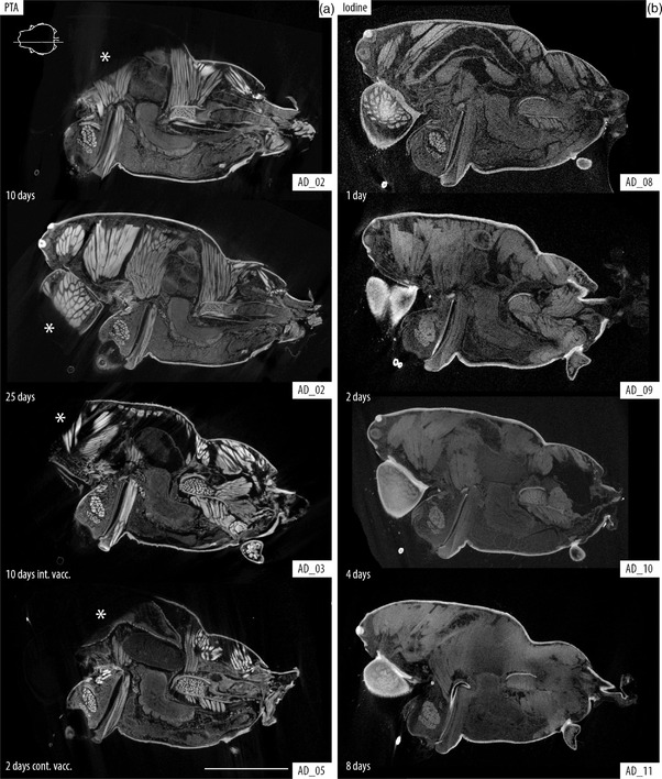
FIGURE 1.

Comparison between PTA and iodine tissue staining. Sagittal slices of the cephalothorax of Araneus diadematus, location indicated with line drawing inset showing cephalothorax in dorsal view. (a) PTA staining after 10 and 25 days of passive diffusion compared with 10 days intermittent and 2 days continuous vacuum. (b) Iodine staining after 1, 2, 4, and 8 days of passive diffusion. Note that although PTA offers a better definition of the structures, there is evident understaining in the cephalic regions and chelicerae (*). Iodine offers better overall staining after 1 day (granted with less definition than PTA); longer times lead to overstaining of the tissues producing a blurry and dull image that lacks definition to discern internal organs. Scale bar: 2 mm
