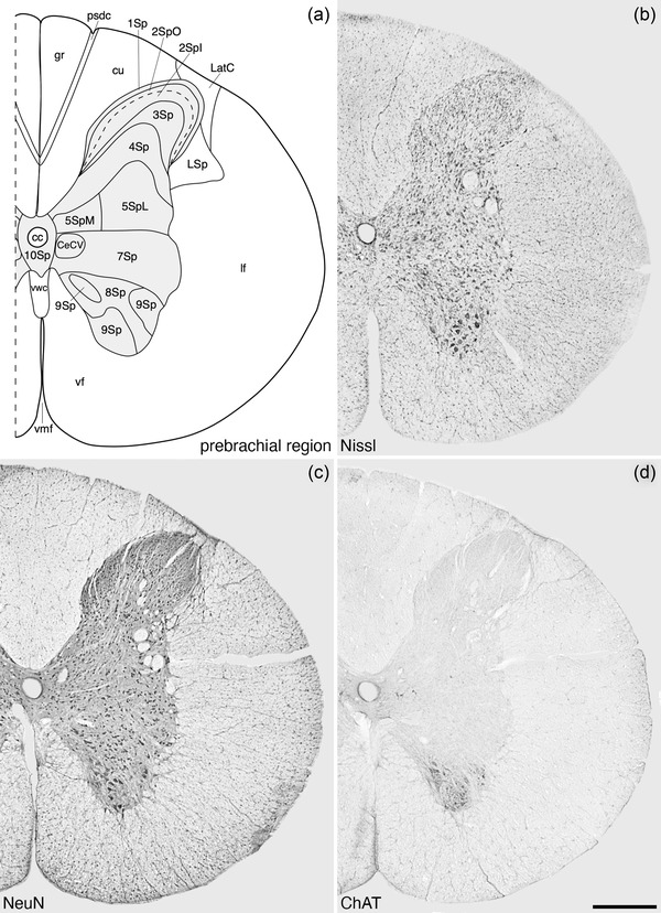
FIGURE 4.

Architectonic reconstruction (a) and low magnification photomicrographs stained for Nissl (b), neuronal nuclear marker (NeuN, c), and choline acetyltransferase (ChAT, d) of coronal sections through the prebrachial region of the tree pangolin spinal cord. In all images, dorsal to the top and medial to the left and correspond to level 2 depicted in Figures 2 and 3. Scale bar in (d) = 500 μm and applies to all images. See list for abbreviations
