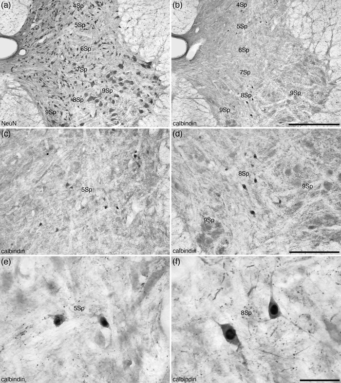
FIGURE 11.

Photomicrographs of coronal sections through the dorsal and ventral horns of the tree pangolin spinal cord stained for neuronal nuclear marker (NeuN, a) and calbindin (b–f) at different magnifications showing the location of calbindin‐immunopositive neurons in laminae 4–9 (Sp4, Sp5, Sp6, Sp7, Sp8, and Sp9) of the spinal gray corresponding to level 4 depicted in Figures 2 and 3. Note that while calbindin‐immunopositive neurons are found in 4Sp, 5Sp, 7Sp, and 8Sp, those in 8Sp (f) are significantly larger than those observed in the other laminae in which they are present, such as Sp5 (e). In all images, dorsal is to the top and medial is to the left. Scale bar in (b) = 500 μm and applies to (a) and (b). Scale bar in (d) = 250 μm and applies to (c) and (d). Scale bar in (f) = 50 μm and applies to (e) and (f). See list for abbreviations
