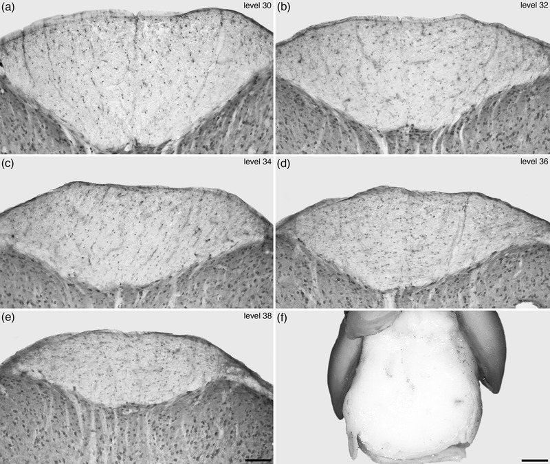
FIGURE 15.

Photomicrographs (a–e) of coronal sections through the spinal cord of the tree pangolin stained for neuronal nuclear marker (NeuN) showing the appearance of the dorsal funiculus in the caudal region of the spinal cord, from levels 30, 32, 34, 36, and 38 depicted in Figures 2 and 3. At levels 30 and 32 the distinct bilaterality of the dorsal funiculus is evident, however, in the more caudal levels (34, 36, 38) this bilaterality becomes increasingly obscured, being faintly visible at level 34 (see also Figure 9d), but then not visible at levels 36 and 38. (f) Photograph of a glabrous skin pad located at the very tip of the ventral aspect of the tree pangolin tail, presumably used for sensory feedback during manipulation and movement of the semi‐prehensile tail. The lack of distinct bilaterality in the most caudal aspects of the dorsal funiculus in the tree pangolin spinal cord may relate to the presence of this glabrous pad. In images (a–e), dorsal is to the top. In image (f) rostral is to the top. Scale bar in (e) = 100 μm and applies to (a–e). Scale bar in (f) = 2 mm
