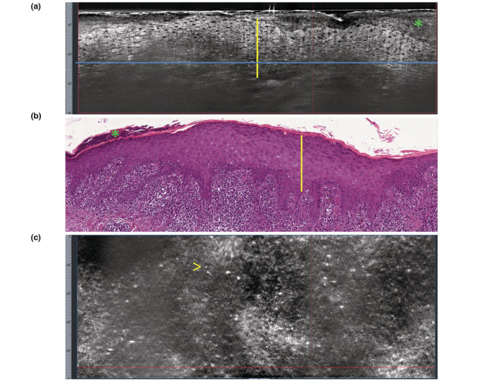
Figure 4.

Lichen planus. (a) Vertical LC‐OCT section shows thickening of the stratum corneum (green asterisk); epidermal thickening (yellow vertical line); poorly defined dermo‐epidermal junction (blue horizontal line). (b) Vertical histopathology correlation shows: hyperorthokeratosis (green asterisk); acanthosis with hypergranulosis (yellow vertical line); ‘band‐like’ lymphohistiocytic infiltrate of the dermo‐epidermal junction and rete ridges sawtoothing (H&E, ×100). (c) Horizontal LC‐OCT section at the level of the dermo‐epidermal junction (taken at the level of the blue line in a) shows absence of evident dermal papillae and presence of small, bright inflammatory cells (arrow).
