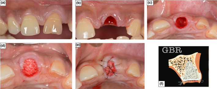
FIGURE 1.

Photographs demonstrating Surgical Protocol for ARP using GBR technique (a) The incisor in position 21 prior to extraction. (b) Atraumatic tooth extraction following incision of the gingival tissue. (c) De‐epithelialization of the gingival tissue collar and localised flap raised. (d) Socket filled with a xenograft bone substitute. (e) The collagen membrane was sutured in place to seal the socket aperture. (f) Graphical representation of ARP using GBR
