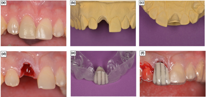
FIGURE 4.

Manufacture of radiographic Measurement stent at extraction site. (a) The incisor in position 11 prior to extraction. (b) 11 sectioned from the cast and buccal aspect trimmed to gingival margin position (c) Palatal aspect trimmed to gingival margin contour. (d) Extraction socket immediately following tooth removal. (e) Radiographic reference stent constructed to marked gingival contour. (f) Gingival margin positional change, immediately following tooth removal
