
FIGURE 12.
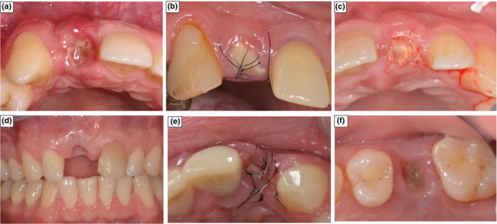
Pictures of socket healing at 2‐weeks demonstrating local Complications. (a) Colour change with GBR group. (b) Dehiscence of the membrane with SS. (c) Dehiscence of the membrane with GBR. (d) Tissue recession with Control. (e) Sequestration of graft with SS. (f) Loss of the membrane with SS