
FIGURE 1.
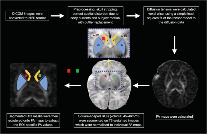
Preprocessing and region‐of‐interest (ROI) analysis of diffusion data. Square‐shaped ROIs (red square: right caudate head; green square: left caudate head) were placed at the centre of the caudate heads. At the level of the basal ganglia/striatum, the caudate heads were defined laterally to the frontal horns of the lateral ventricles (LV) and bounded postero‐laterally by the anterior limb of the internal capsule (ALIC; purple line). The ALIC is an easily identifiable white matter tract on FA maps, seen as a high‐intensity structure (due to its high anisotropy) between the lentiform nucleus (LN) and caudate head