
FIGURE 6.
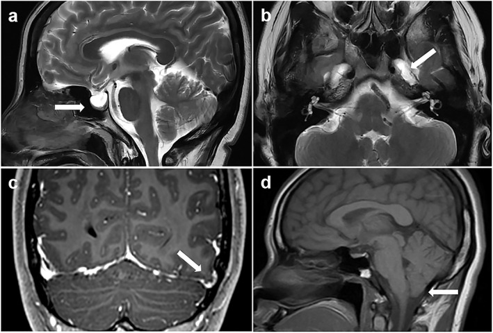
Classical magnetic resonance imaging (MRI) signs suggestive of elevated intracranial pressure. (a) Sagittal T2‐weighted MRI showing an empty sella (arrow). (b) Axial T2‐weighted MRI showing the enlarged Meckel's caves bilaterally (white arrow points to left Meckel's cave). (c) Coronal T1‐weighted postcontrast MRI showing a left temporal lobe cephalocele (arrow). (d) Sagittal T1‐weighted MRI showing mild inferior cerebellar tonsillar descent (arrow).