
FIGURE 7.
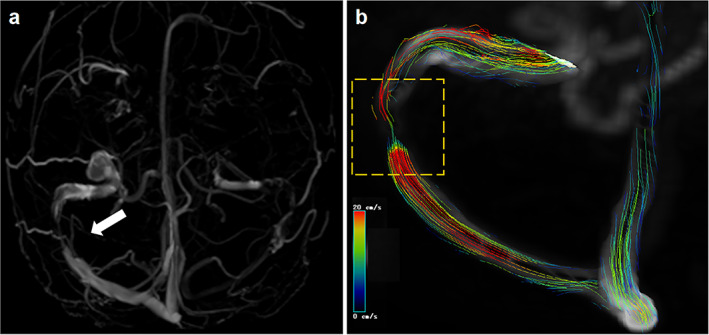
Four‐dimensional (4D) flow magnetic resonance imaging (MRI) showing the hemodynamic of cerebral venous sinus stenosis. A female presented with severe headache and papilloedema with a clinical diagnosis of idiopathic intracranial hypertension (IIH). (a) Inferior reconstruction of magnetic resonance venography (MRV) showed the drainage dominance of right transverse sinus and right transverse sinus stenosis. (b) 4D flow pathlines indicate abnormal flow condition and the existence of stenosis. The color coding of the pathlines reflects the magnitude of blood flow velocities in the vasculature (blue = low, red = high). The white arrow and yellow‐dashed square in the artwork mean the transverse sinus stenosis.