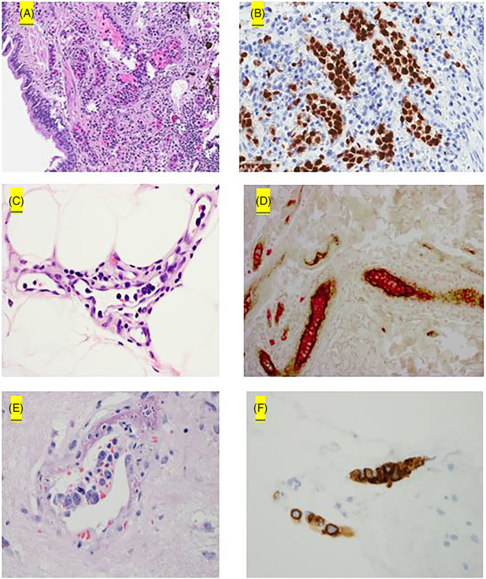
FIGURE 2.

Pathologic findings in IVL. (A, B) IVL in the lung is highlighted by PAX5. (C, D) IVL in the skin with lymphoma cells positive for CD20 (red) within the vessels and vessel wall highlighted by Factor VIII (brown). (E, F) CNS‐IVL with lymphoma cells the IVL cells positive for CD20 [Color figure can be viewed at wileyonlinelibrary.com]
