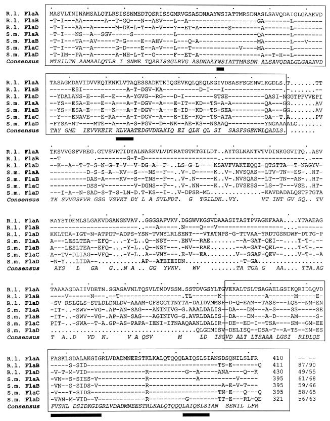
FIG. 2.
Comparison of the three R. lupini (R.l.) and four S. meliloti (S.m.) deduced flagellin polypeptide sequences. Numbering refers to amino acid residues in each line. Dashes signify identical residues with respect to the R. lupini FlaA sequence, and dots signify gaps. The consensus sequence includes all residues that form a homology group with a weighted relative frequency of 0.5 or greater. Conserved N- and C-terminal domains are boxed, and black bars denote amino acid residuces unique to right-handed helical flagellar filaments (41). Identity/similarity values (percentages) relative to R. lupini FlaA are listed at the end of each sequence.

