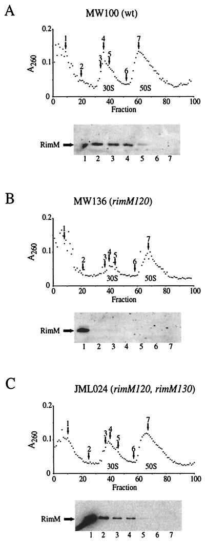
FIG. 2.
Cellular localization of RimM proteins in the wild type (A), rimM120 mutant (B), and rimM120 mutant overexpressing RimM (C). Cell extracts were fractionated by sucrose gradient centrifugation during conditions that dissociated the 70S ribosomes into 50S and 30S subunits. Selected fractions, indicated by arrows above the A260 curve, were screened for the presence of the RimM proteins in Western blotting experiments using a polyclonal anti-RimM antiserum. The locations of the 50S and 30S ribosomal subunits are indicated below the A260 curve.

