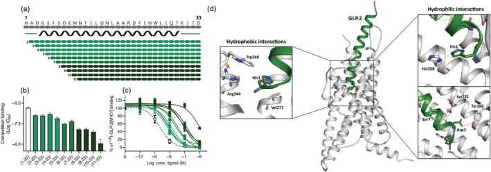
FIGURE 1.

N‐terminally truncated GLP‐2 variants on the human GLP‐2R. (a) Schematic overview of the N‐terminally truncated GLP‐2 variants. The black spiral indicates the predicted α‐helix structure from amino acid number 4 to amino acid number 29 (34). COS‐7 cells were transiently transfected with the human GLP‐2R and the N‐terminally truncated GLP‐2 variants competed with [125I]‐GLP‐2(1‐33)[M10Y] giving the (b) log IC50 values calculated from (c) the inhibition curves. The dashed line in (c) represents human GLP‐2. Data are shown as mean ± SEM, from n = 3 independent experiments carried out in duplicate. (d) A representation of the human GLP‐2R structure (grey—PDBid: 7D68) in complex with the full length GLP‐2 peptide (green) is showed where the interactions between the N‐terminal GLP‐2 residues and the GLP‐2R are highlighted.
