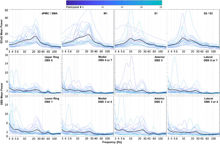
FIG 2.

Cortical and subcortical field potentials at rest in participants with Parkinson's disease. Spectral power from all electrode contacts over the ipsilateral cortex (first row) and the subthalamic nucleus (STN) region (second and third rows) are categorized by anatomical location obtained from imaging, regardless of dystonia status. For each location, there is one plot per participant color‐coded by participant number. Means are represented by darker black lines. Beta frequency power is present and relatively large in essentially all electrocorticography (ECoG) contacts, whereas lower beta power is present in most, but not all, deep brain stimulation (DBS) contacts, along with greater relative power at lower frequencies. dPMC, dorsal premotor cortex; M1, primary motor cortex; S1, primary somatosensory cortex; SMA, supplementary motor area.
