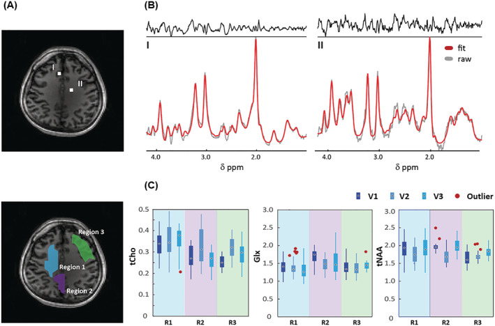
FIGURE 4.

(A) Anatomical brain image with two voxel locations (top) and the three different regions of interest (ROIs) (bottom). (B) MR spectra obtained with crusher coil and L2‐regularization of the two voxels in (A) with LCModel fit. (C) Concentration ratios (tCho, Glx, and tNAA) to total Cr (tCr) as calculated by LCModel for each ROI. The number of voxels of these ROIs (R1/R2/R3) in a matrix size of 38 × 38 is 48/16/38 for volunteer 1 (V1), 52/25/26 for volunteer 2 (V2), and 34/29/27 for volunteer 3 (V3) (see Figure S2)
