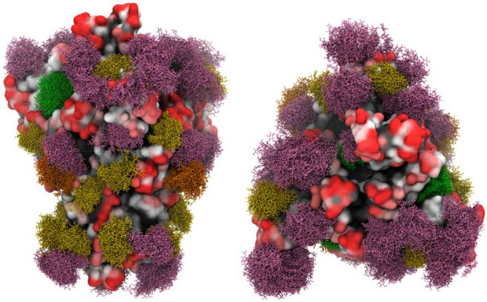
Fig. 15.

3D models of the glycosylated S (spike) protein of SARS‐CoV‐2. High‐mannose structures Man9 are depicted in green and Man5 in dark yellow. Hybrid N‐glycans are shown in orange and complex N‐glycans in pink. Figure reproduced from Ref. [321].
