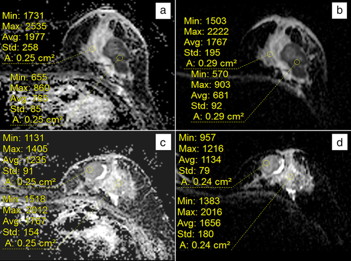
FIGURE 1.

Representative ADC images of RS‐EPI and SS‐EPI sequences from a breast cancer patient with HER‐2‐negative and HER‐2‐positive invasive ductal carcinoma; (a) and (b) are HER‐2‐negative images of a left breast invasive ductal carcinoma patient. Measuring the ADC map revealed a mean ADC value of 0.765 × 10⁻³ mm2/sec by RS‐EPI (a) and 0.681 × 10⁻³ mm2/sec by SS‐EPI (b); (c) and (d) are images of a HER‐2‐positive left breast invasive ductal carcinoma patient. Measuring the ADC map revealed a mean ADC value of 1.235 × 10⁻³ mm2/sec by RS‐EPI (c) and 1.134 × 10⁻³ mm2/sec by SS‐EPI (d).
