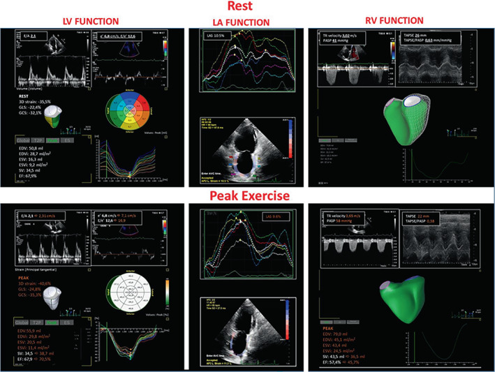
Figure 7.

Cardiopulmonary exercise test imaging rest to peak exercise analysis of the same case as Figure 5 . Measures obtained by stress echocardiography (rest to peak exercise). The analysis was performed analysing the diastolic (E/e') and systolic (three‐dimensional longitudinal and circumferential strain) left ventricular (LV) function; the adaptive left atrial (LA) dynamics by LA strain (LAS); right ventricular (RV) function (RV ejection fraction 3D analysis) and its coupling with the pulmonary circulation by the tricuspid annular plane systolic excursion/systolic pulmonary artery pressure (TAPSE/PASP) ratio. Data are reported at rest (white) and at peak exercise (orange) with the changes occurring in the main variables from rest to peak. TR, tricuspid regurgitation.
