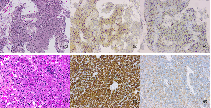
FIGURE 2.

Histotypes of corticotroph aggressive tumors. (A) Silent corticotroph tumors are typically composed of regular cells with chromophobic or slightly basophilic cytoplasm (hematoxylin, phloxine, saffron [HPS] staining, original magnification [OM] × 200). (B) Silent corticotroph tumors express the T‐PIT transcription factor as do all other tumors of the corticotroph lineage (T‐PIT immunohistochemistry, OM × 100). (C) Some silent corticotroph tumors show immunoreactivity for GATA3, a transcription factor also expressed in gonadotroph tumors (GATA3 immunohistochemistry, OM × 100). (D) Crooke cell adenoma is composed of neoplastic cells harboring the ring‐like hyaline change typical of normal corticotroph cells of patients with hypercortisolism (HPS staining, OM × 200). (E) The ring‐like hyaline material corresponds to an accumulation of low‐molecular‐weight cytokeratins (cytokeratin 18 immunohistochemistry, OM × 200). (F) Expression of ACTH in Crooke cell adenomas is typically restricted to the paranuclear and peripheral regions of the cytoplasm (ACTH immunohistochemistry, OM × 200).
