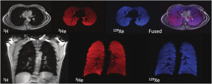
FIGURE 6.

MR images of 1H (grey), 3He (red) and 129Xe (blue) acquired from a healthy volunteer in the same breath‐hold containing 600 mL of 129Xe and 300 mL of 3He. The anatomic 1H images show excellent spatial registration with the 3He and 129Xe ventilation images, as demonstrated by the overlaid fused image (purple). Figure reproduced with permission from Reference 37
