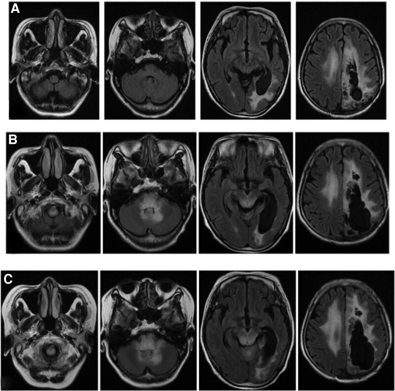
Figure 3.
The T2 flair sequence of MRI. (1) Post-operation (A), this admission (B) and follow-up after discharge (C); (2) compared with post-operation, the brain stem and cerebellum developed diffuse hyperintensity at this admission; (3) compared with this admission, the brain stem and cerebellum developed more diffuse hyperintensity during follow-up.

