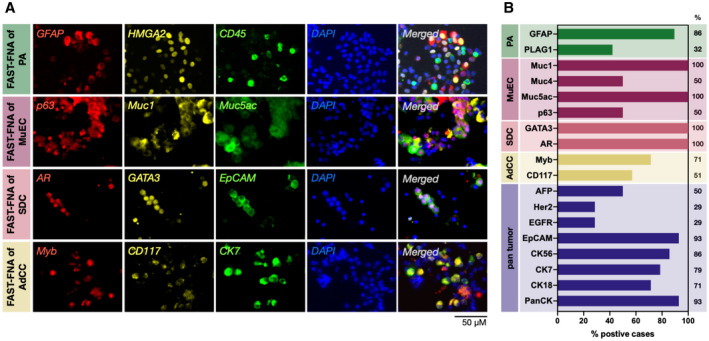
Figure 2.

Representative examples of FAST‐fine‐needle aspiration (FNA) analysis. (A) Single‐cell staining with a panel of salivary gland tumor (SGT)‐relevant antibodies (pleomorphic adenoma [PA], mucoepidermoid carcinoma [MuEC], salivary duct carcinoma [SDC], adenoid cystic carcinoma [AdCC]). Antibodies were conjugated to FAST probes with one of the following fluorophores: Alexa fluor 488, Alexa fluor 555, or Alexa fluor 647. (B) FAST‐FNA analysis of SGT subtypes (PA, n = 19; MuEC, n = 3; SDC, n = 4; AdCC, n = 6; all malignant tumors n = 24) showing high expression levels of different biomarkers of each SGT subtype. Sample positivity was determined if more than 20% of tumor cells expressed the respective marker. The percentages refer to the number of patients in whom a given biomarker was positive.
