FIGURE 1.

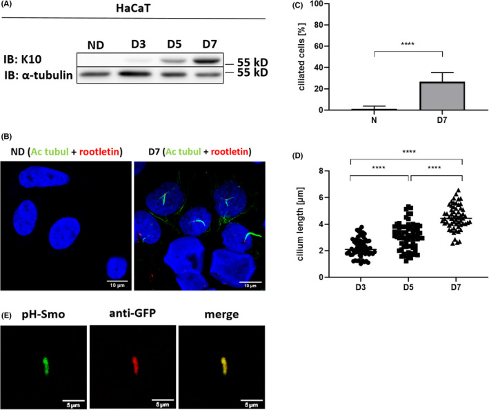
Differentiation of HaCaT keratinocytes increases their ciliation. (A) Western blot showing a representative image of K10 protein expression during differentiation of HaCaT. α‐tubulin used as a loading control. (B) Representative immunofluorescence images of acetylated tubulin (green) and rootletin (red) co‐staining in non‐differentiated and 7 days‐differentiated HaCaT. Scale bar, 10 μm. (C) Quantification of ciliated HaCaT upon non‐differentiated and 7 days differentiated conditions. Unpaired t test with Welch's correction (N = 10, 16). (D) Cilium length measured in HaCaT at D3, D5 and D7 of differentiation. Ordinary one‐way ANOVA test with Holm‐Šidák's correction (N = 64, 68, 59). (E) Representative images of SMO IN/OUT assay in 7 days‐differentiated HaCaT. Scale bar, 5 μm. Ac‐tubul, acectylated tubulin; IB, immunoblot; ND, non‐differentiated, D3, D5, D7: day 3, 5 and 7 of differentiation, respectively
