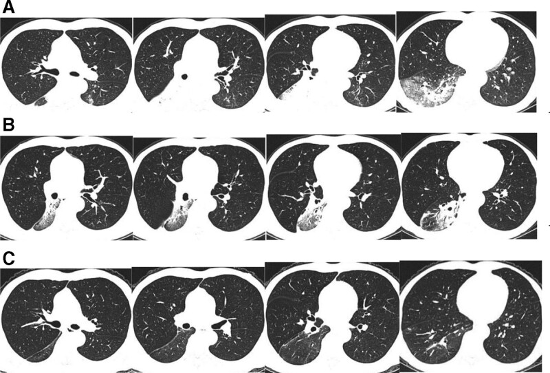
Figure 4.
Computed tomography. A: There was a large consolidation in the dorsal segment of the right lower lobe, patchy exudation with partial consolidation in the basal segment of the right lower lobe, and right lower hilar lymphadenopathy (December 7, 2020); B: The lesion was obviously absorbed (January 4, 2021); C: The lesion was almost completely absorbed with few fibrous cords remaining (February 15, 2021).

