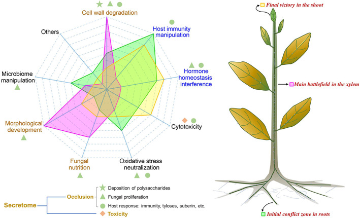
Fig. 4.

Intensity model of biological functions of the secretome at different stages of Verticillium wilt infection. Left: the main contributions of different functions of the secretome in polysaccharide deposition, fungal proliferation, host response and toxicity. Cell wall degradation, morphological development and fungal nutrition predation (shown in brown font) represent the main effects of the secretome in vessels; and host immunity manipulation and hormone interference (shown in blue font) represent the main effects of the secretome in roots and leaves. The green region shows the biological functions of the secretome that operate during the initial stages of the colonization, the pink region shows those operating when the fungus is present in the xylem, and the yellow region is for the final stage of infection when the pathogen has reached the leaves (see plant image on right).
